
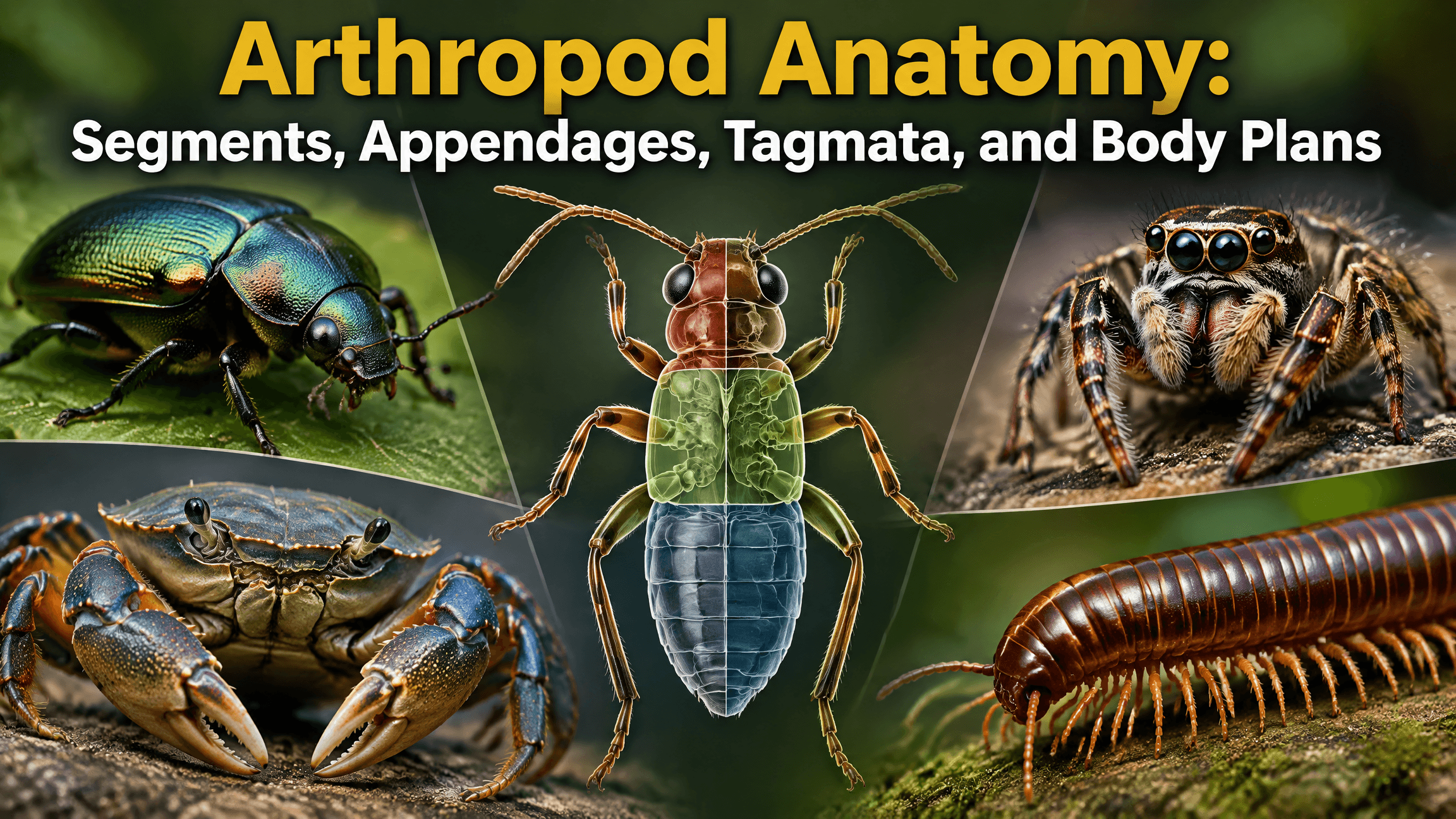
Arthropod anatomy is built around a modular body plan. The body is fundamentally segmented, paired jointed appendages are associated with segments, and the outside is covered by a cuticle that functions as an exoskeletal system. Over evolutionary time, those segments and appendages have been grouped, fused, reduced, and specialized into remarkably different forms.
That is why a beetle, spider, crab, centipede, and millipede can all be arthropods without sharing the same visible body regions or number of walking legs. Their anatomy follows the same broad construction principles, but each lineage reorganizes those principles differently.
The key ideas are segmentation, appendage specialization, and tagmosis. Tagmosis is the grouping of segments into larger functional regions called tagmata. Insects typically have a head, thorax, and abdomen. Spiders have a prosoma and opisthosoma. Myriapods generally have a head and long trunk. Crustacean body regions are especially variable.
Quick Overview of Arthropod Anatomy
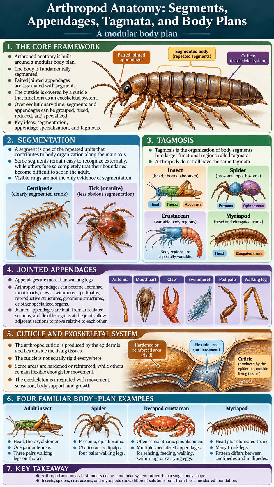
The Smithsonian overview of the arthropod body plan describes arthropods as bilaterally organized, typically segmented animals with an external skeleton and specialized appendages. Those appendages can serve in feeding, respiration, reproduction, walking, swimming, and other functions. These anatomical variations help produce the broader diversity of arthropod body plans and adaptations.
This modular organization is the central idea behind arthropod anatomy. Instead of one body shape being copied across the entire phylum, repeated units can be modified for different jobs. One appendage may remain a walking leg, another may become a mouthpart, and another may become a sensory or reproductive structure. Segmentation, jointed appendages, the cuticle, and tagmosis are central to the defining arthropod traits.
Visible anatomy can therefore look very different among groups even when the deeper construction plan is related. The anatomy of an arthropod makes the most sense when body regions and appendages are studied together rather than as isolated parts.
Segmentation: The Foundation of the Arthropod Body Plan
What a Body Segment Is
A segment is one of the repeated units that contributes to the organization of the body along its main axis. In an idealized segmented animal, multiple units repeat one after another. Real arthropods modify that arrangement extensively.
Segments may differ in size, shape, appendages, internal organs, or function. Some remain easy to recognize externally, while others fuse so completely that their boundaries become difficult to see in the adult.
A centipede makes segmentation obvious because much of its trunk displays a repeated sequence of leg-bearing units. A spider or tick shows how heavily the same basic principle can be reorganized.
Visible Segments Versus Ancestral Segmentation
Not every arthropod looks obviously segmented. Fusion can blur or erase external boundaries, but that does not mean segmentation disappeared from the evolutionary body plan.
Embryology, nerves, muscles, appendage positions, internal structures, and comparative anatomy can reveal segmental organization that is difficult to see on the surface. This is especially important in compact-bodied arthropods such as mites and ticks.
Visible rings are therefore not the only evidence of segmentation. An adult body can be highly integrated while retaining a developmental and anatomical history built from segments.
Tagmosis: How Segments Become Functional Body Regions
What Tagmosis Means
Tagmosis is the organization of body segments into larger functional regions called tagmata. A tagma is not simply a decorative body division. It reflects coordination among neighboring segments for related tasks.
For example, a region can concentrate sensory structures and feeding appendages, while another carries locomotor appendages. Other regions may emphasize digestion, reproduction, respiration, or locomotion.
Tagmosis is one of the main ways arthropods turn a repeated segmented framework into an integrated animal. Different lineages solve this problem differently.
Why Arthropods Do Not All Have the Same Tagmata
There is no universal arthropod arrangement equivalent to “head, thorax, abdomen.” That pattern belongs to insects. Other arthropod lineages organize segments in different combinations.
Spiders combine the anterior body into a prosoma and follow it with an opisthosoma. Many crustaceans integrate head and thoracic segments into a cephalothorax, but crustacean tagmosis is too diverse for one formula. Myriapods generally retain an obvious head followed by an elongated trunk.
The useful rule is that arthropods group and specialize segments, not that all arthropods use the same named regions.
Jointed Appendages and Why They Matter
Appendages Are More Than Walking Legs
The word “appendage” is broader than “leg.” Arthropod appendages can become antennae, mouthparts, claws, swimmerets, pedipalps, reproductive structures, grooming structures, or other specialized organs.
The Smithsonian describes appendage specialization as a major feature of the arthropod body plan. A modular system allows structures associated with particular segments to evolve new functions without redesigning the entire animal.
This helps explain why arthropods can occupy such different ecological roles. A basic appendage framework can be altered for sensing, grasping, chewing, swimming, digging, mating, carrying eggs, or walking. Jointed limbs and exoskeletal leverage provide the mechanical foundation for arthropod locomotion.
How Joints Create Controlled Movement
Jointed appendages are built from articulated sections rather than one continuous rigid rod. Flexible regions at the joints allow adjacent sections to move relative to each other.
Muscles act across these joints, and the external skeletal system provides surfaces against which those muscles can work. This arrangement gives arthropods precise control over limbs and mouthparts.
The exact mechanics vary. A crustacean claw, insect leg, spider pedipalp, and centipede walking leg do not all move in the same way, but each reflects a repeated jointed architecture.
The Arthropod Cuticle and Exoskeletal System
Cuticle, Chitin, and Protein
The arthropod cuticle is produced by the epidermis and lies outside the living tissues. Smithsonian describes the exoskeleton as containing chitin embedded in a protein matrix. The material properties vary from one body region and lineage to another.
Some areas are hardened or reinforced, while others remain flexible enough for movement. A joint cannot function if every part of the covering is equally rigid.
Crustaceans may additionally mineralize parts of the cuticle, often with calcium salts. That creates the strongly reinforced body coverings familiar in many crabs and lobsters, but mineralization is not a universal feature of all arthropods.
Why the Exoskeleton Is Not a Separate Suit of Armor
It is easy to imagine the exoskeleton as a shell worn by the animal, but that image is incomplete. Living epidermal tissue produces the cuticle, muscles attach to the inner surfaces of the exoskeletal system, and sensory structures pass through or interact with it.
The outer covering is therefore integrated with movement, sensation, body support, and growth. Its biology is inseparable from the tissues underneath.
The exoskeleton also has to be replaced as the animal grows, which is why molting is so closely connected to arthropod anatomy. The details of that growth process belong to the molt cycle rather than to anatomy alone.
Insect Body Plan: Head, Thorax, and Abdomen
The Head
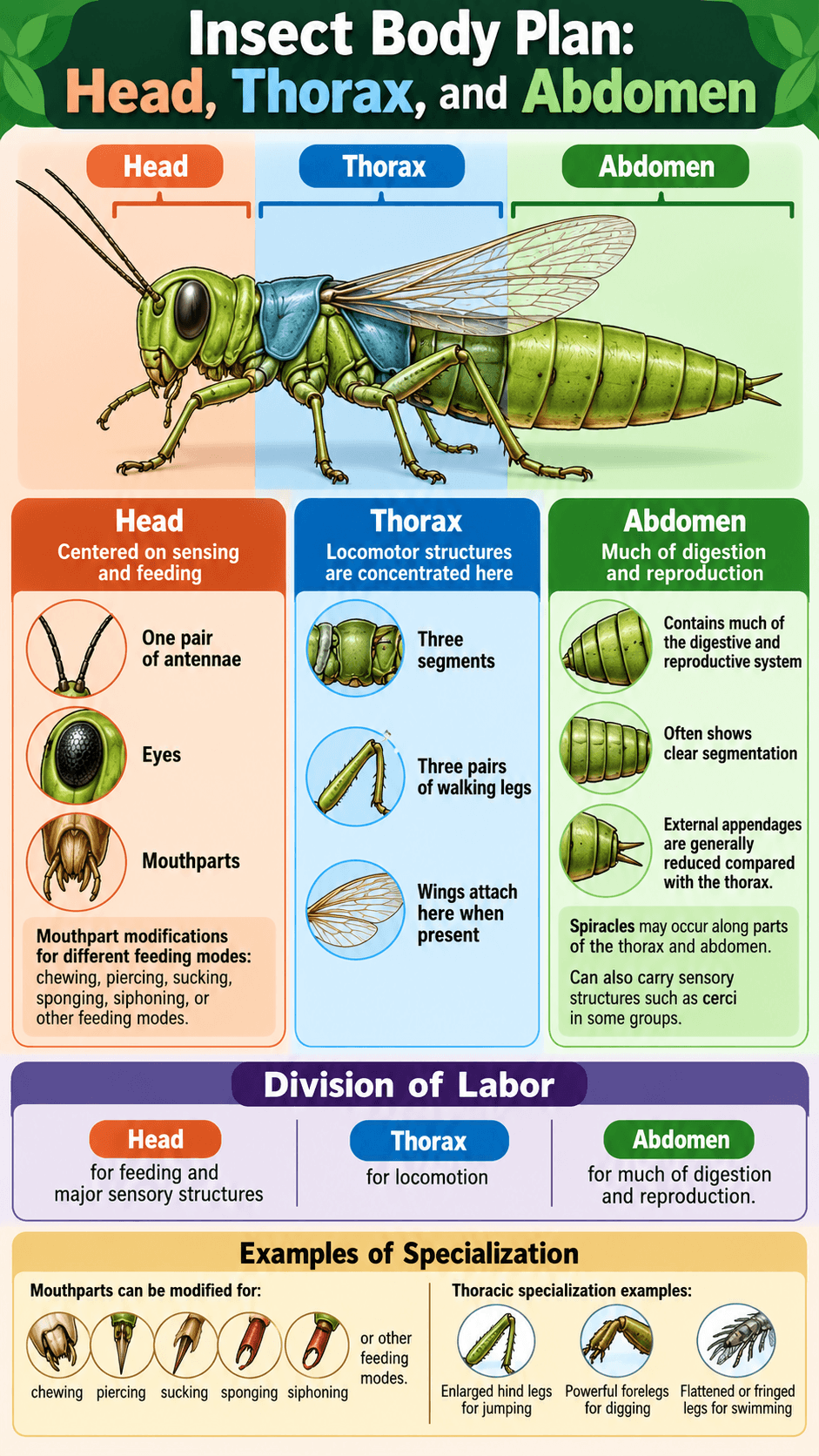
The insect head concentrates many sensory and feeding structures. It typically carries one pair of antennae, eyes, and mouthparts. The exact mouthpart arrangement varies widely among insects because the ancestral components can be modified for chewing, piercing, sucking, sponging, siphoning, or other feeding modes.
The head is therefore an excellent example of tagmosis. Multiple ancestral segments and appendages are integrated into one compact region centered on sensing and feeding.
The Thorax
The thorax consists of three segments and bears the insect’s three pairs of walking legs. The University of Kentucky insect anatomy guide identifies the head, thorax, and abdomen as the basic insect body regions and places the three pairs of legs on the thorax.
When wings are present, they also attach to the thorax. This concentrates most adult insect locomotor structures in one tagma.
Even within this fixed framework, thoracic structures can be highly specialized. Jumping insects may have enlarged hind legs, digging insects may have powerful forelegs, and swimming insects may flatten or fringe their legs for movement through water.
The Abdomen
The insect abdomen contains much of the digestive and reproductive system and often shows clear segmentation. External appendages are generally reduced compared with the thorax, although terminal or reproductive structures can be important.
Spiracles may occur along parts of the thorax and abdomen in terrestrial insects, connecting the outside air with the tracheal respiratory system. The abdomen can also carry sensory structures such as cerci in some groups.
The insect pattern shows a strong division of labor: head for feeding and major sensory structures, thorax for locomotion, and abdomen for much of digestion and reproduction. Real species modify that pattern without losing the overall three-region arrangement.
Spider and Arachnid Body Plans
Prosoma and Opisthosoma
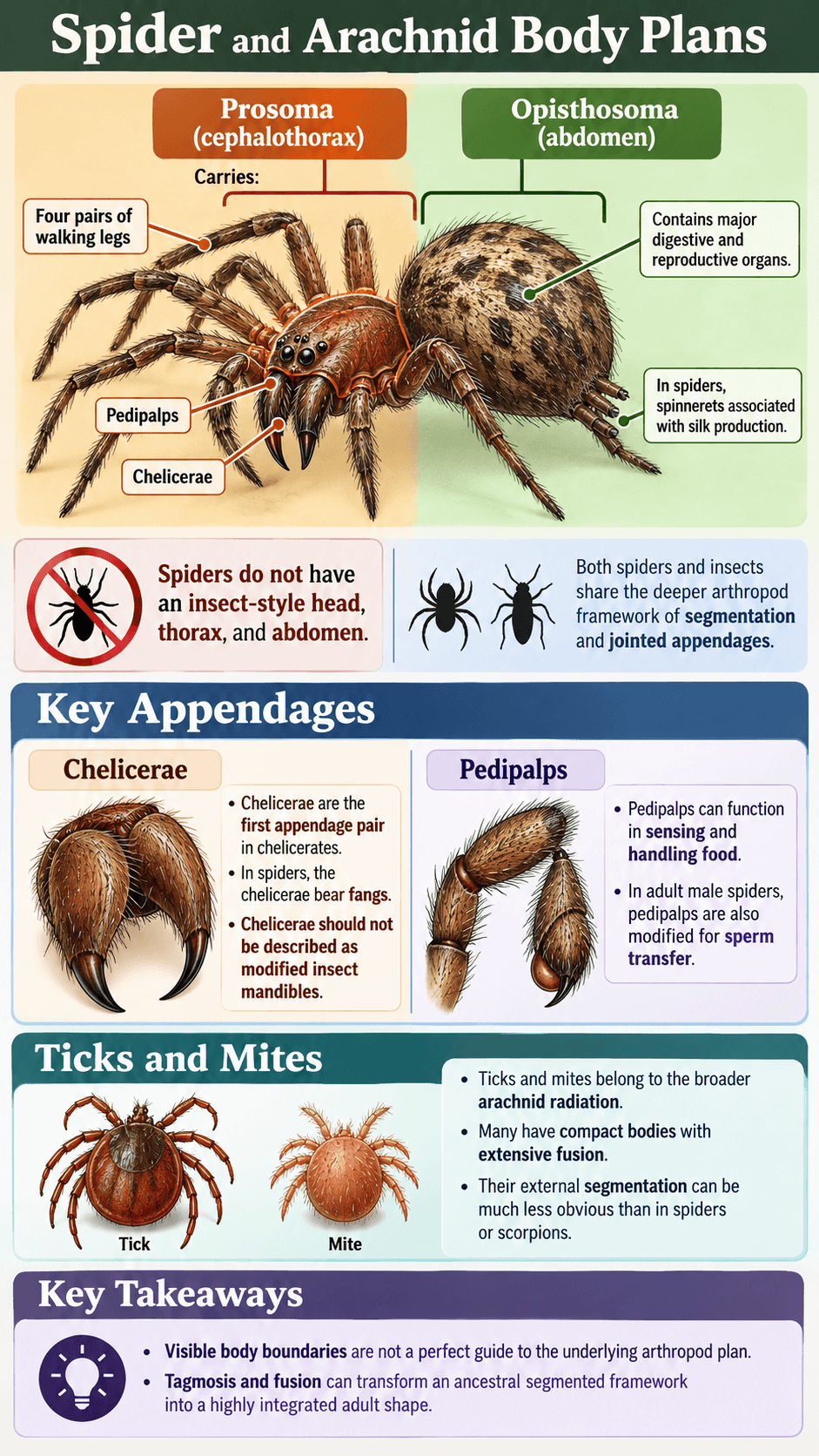
Spiders do not have an insect-style head, thorax, and abdomen. Their anterior body region is the prosoma, often called the cephalothorax, and the posterior region is the opisthosoma, commonly called the abdomen.
The prosoma carries the chelicerae, pedipalps, and four pairs of walking legs. The opisthosoma contains major digestive and reproductive organs and, in spiders, the spinnerets associated with silk production.
This organization immediately distinguishes spider anatomy from insect anatomy even though both animals share the deeper arthropod framework of segmentation and jointed appendages.
Chelicerae and Pedipalps
Chelicerae are the first appendage pair in chelicerates. In spiders, the chelicerae bear fangs. They should not be described as modified insect mandibles because chelicerates and mandibulate arthropods follow different appendage histories.
Pedipalps form the next appendage pair. They can function in sensing and handling food, and in adult male spiders they are also modified for sperm transfer.
The Smithsonian tarantula resource notes that spider fangs occur at the tips of the chelicerae, illustrating how chelicerate mouth-area appendages differ from insect mandibles.
Why Ticks and Mites Can Look Less Segmented
Ticks and mites belong to the broader arachnid radiation, but many have compact bodies with extensive fusion. Their external segmentation can be much less obvious than in spiders or scorpions.
This is a useful reminder that visible body boundaries are not a perfect guide to the underlying arthropod plan. Tagmosis and fusion can transform an ancestral segmented framework into a highly integrated adult shape.
Crustacean Body Plans Are Highly Variable

Why There Is No Single Crustacean Formula
Crustaceans include crabs, shrimp, lobsters, crayfish, copepods, amphipods, isopods, barnacles, ostracods, branchiopods, krill, and many other forms. Their body regions and appendage patterns vary far more than a simple crab model suggests.
The Smithsonian’s reference work on crustacean functional morphology and appendage diversity highlights topics including carapace structure, antennules, antennae, walking, swimming, burrowing, grooming, and reproduction. That breadth reflects how extensively crustacean appendages are specialized.
Some groups have a conspicuous carapace covering fused regions, while others have exposed body segments. Appendages can be biramous or modified in many ways, and not every crustacean follows the decapod pattern familiar from crabs and lobsters.
Decapod Cephalothorax and Abdomen
In many decapod crustaceans, head and thoracic segments are functionally integrated into a cephalothorax. A carapace often covers much of this region. The abdomen follows behind and varies from muscular and mobile in shrimp and lobsters to folded beneath the body in true crabs.
The cephalothorax carries sensory appendages, mouthparts, and thoracic limbs. Some of those thoracic limbs may become claws, while others function primarily in walking.
This organization is useful for understanding decapods, but it should not be generalized to all crustaceans.
Barnacles Show Extreme Modification
Barnacles are crustaceans despite looking very unlike a shrimp or crab as adults. They attach to surfaces and surround much of the body with protective plates. Their feeding appendages extend into the water to capture suspended particles.
The Smithsonian identifies goose barnacles within Arthropoda and Crustacea, making them a strong example of how far an arthropod body plan can be modified while remaining recognizable through developmental and anatomical evidence.
Myriapod Body Plans
Head and Elongated Trunk
Myriapods generally have a distinct head followed by an elongated trunk made of many segments. The repeated trunk pattern makes segmentation especially easy to see.
The head bears sensory and feeding structures, while much of the trunk is associated with locomotion. The exact arrangement differs among centipedes, millipedes, pauropods, and symphylans.
Because the trunk repeats many similar units, myriapods provide one of the clearest living examples of the modular basis of arthropod anatomy.
Centipede Trunk Organization
Centipedes typically have one pair of walking legs on each leg-bearing trunk segment. The first trunk appendages are modified into forcipules, structures used in prey capture and venom delivery.
This demonstrates how appendages associated with a particular segment can shift from locomotion to feeding. The rest of the trunk retains a more obviously repeated walking-leg pattern.
Centipede anatomy should not be summarized as “many identical leg segments” because the anterior trunk is strongly specialized.
Millipede Diplosegments
Millipede anatomy follows a different trunk pattern. Many apparent trunk rings are diplosegments, units formed through developmental fusion that typically bear two pairs of legs.
That distinction is why millipedes can have a dense sequence of legs without simply being centipedes with extra limbs. Their segment organization is developmentally different.
The difference also provides a useful visual lesson in tagmosis and fusion: adult external units do not always correspond one-to-one with ancestral segments.
Appendage Homology and Specialization
What Homology Means
Homologous structures are related through common ancestry even if their modern functions differ. Arthropod appendages are a powerful example because ancestral segmental appendages have been repeatedly modified in different lineages.
Homology does not mean every mouthpart across Arthropoda is the same structure. Comparisons must be made carefully within the correct evolutionary framework.
For example, a spider chelicera should not simply be labeled the equivalent of an insect mandible. Both participate in feeding, but functional similarity does not establish direct one-to-one homology.
From Locomotion to Feeding, Sensing, and Reproduction
Appendages can shift from one role to another over evolutionary time. Antennae emphasize sensing. Mandibles and maxillae participate in feeding. Crustacean swimmerets may aid swimming, reproduction, or egg carrying depending on group and sex.
Pedipalps can assist with sensation, prey handling, or reproduction. Claws may evolve from walking appendages and become structures for grasping, defense, display, or food handling.
The modularity of the arthropod body allows these changes to occur locally. A lineage can transform one appendage pair without necessarily changing every other limb.
Internal Anatomy Beneath the Exoskeleton

Muscles and the Exoskeletal Framework
Arthropod muscles act against the exoskeletal system. Rather than attaching to internal bones as vertebrate muscles do, many locomotor muscles attach to internal surfaces or projections associated with the cuticle.
When muscles contract, they move articulated parts around joints. This arrangement can produce very fine control despite the external position of the skeleton.
The design also explains why flexible joint membranes are essential. Rigid plates provide leverage and support, while flexible connections permit movement.
Nervous System and Segmental Organization
The arthropod nervous system generally includes a brain in the anterior body and a ventral nerve cord with segmentally organized ganglia or centers. The degree of fusion varies with body organization.
In animals with strongly integrated tagmata, nervous structures can also become concentrated. This parallels the broader anatomical pattern in which repeated segments evolve into coordinated functional regions.
Detailed sensory processing differs dramatically among lineages, so anatomy alone does not predict how an insect, spider, or crustacean perceives its environment.
Open Circulation and the Body Cavity
Arthropods generally use an open circulatory system. Hemolymph leaves vessels and moves through body spaces before returning toward the heart.
The circulatory system interacts differently with respiration in different groups. In many insects, the tracheal system delivers much of the oxygen directly to tissues. In many crustaceans and chelicerates, hemolymph plays a larger role in transporting respiratory gases. Internal body organization also differs alongside the various arthropod respiratory systems.
This variation is important because internal anatomy follows the same general pattern as external anatomy: common structural themes are modified for different lineages and environments.
Why Arthropod Anatomy Is So Variable
Modularity Makes Specialization Possible
A modular body plan allows one part of the animal to become specialized without requiring every other part to change in the same way. Repeated segments provide a framework that evolution can modify locally.
This helps explain the contrast between a streamlined swimming crustacean, a jumping insect, a web-building spider, and a burrowing millipede. Each animal emphasizes different regions and appendages.
Fusion Can Increase Functional Integration
Segment fusion is not simply a loss of complexity. Fusing neighboring units can create a stronger or more coordinated body region.
An insect thorax concentrates locomotor structures. A spider prosoma integrates feeding, sensory, and locomotor appendages. A decapod cephalothorax combines multiple anterior functions under a carapace.
Tagmosis therefore represents functional reorganization rather than merely making segments harder to see.
Reduction Can Be as Important as Addition
Evolutionary specialization can involve loss or reduction as well as enlargement. Appendages may become tiny, disappear externally, fuse with neighboring structures, or stop serving their ancestral role.
Highly specialized parasites, attached crustaceans, and compact mites show how an arthropod can depart dramatically from a familiar walking-body form.
Understanding anatomy requires attention to what has been modified or reduced, not just to obvious new structures.
Common Arthropod Anatomy Mistakes
All Arthropods Have a Head, Thorax, and Abdomen
No. That arrangement is characteristic of insects. Spiders use a prosoma and opisthosoma, many myriapods have a head and trunk, and crustacean tagmata vary substantially.
Every Segment Has a Walking Leg
No. Appendages can become antennae, mouthparts, claws, swimmerets, reproductive structures, or other specialized organs. Some segments may also lose appendages entirely.
The Exoskeleton Is Completely Rigid
No. Arthropods need flexible membranes and articulations at joints. Different parts of the cuticle can have very different mechanical properties.
Spider Fangs Are Modified Insect Mandibles
No. Spider fangs are associated with the chelicerae, while insect mandibles belong to a different appendage system within mandibulate arthropods. Similar feeding roles do not make the structures identical in evolutionary origin.
More Segments Mean a More Primitive Animal
No. Segment number alone does not provide a simple scale from “primitive” to “advanced.” Arthropod lineages have evolved through different patterns of fusion, specialization, reduction, and repetition.
How Body Regions Differ Across Four Familiar Arthropods
| Example | Main body regions | Key appendage pattern | Useful anatomical lesson |
|---|---|---|---|
| Adult insect | Head, thorax, abdomen | One pair antennae; three pairs walking legs on thorax | Strong division between sensory-feeding, locomotor, and abdominal functions |
| Spider | Prosoma, opisthosoma | Chelicerae, pedipalps, four pairs walking legs | Chelicerate organization differs fundamentally from insect anatomy |
| Decapod crustacean | Often cephalothorax plus abdomen | Multiple specialized appendages for sensing, feeding, walking, swimming, or carrying eggs | Crustacean appendages are highly diverse and multifunctional |
| Myriapod | Head plus elongated trunk | Many trunk legs; pattern differs between centipedes and millipedes | Repeated segmentation remains especially visible |
The table is a comparison of familiar forms, not a complete description of every member of each lineage. Arthropod anatomy is most accurate when examples are treated as representative patterns rather than universal templates.
FAQ
What Are the Main Parts of an Arthropod Body?
There is no single set of named body regions for every arthropod. The common structural themes are segmentation, jointed appendages, and an external cuticular skeleton. Segments are often grouped into functional tagmata, but the names and boundaries of those tagmata differ among insects, arachnids, crustaceans, and myriapods.
What Is Tagmosis in Arthropods?
Tagmosis is the grouping and specialization of body segments into larger functional regions called tagmata. Insects show tagmosis as head, thorax, and abdomen, while spiders show a different pattern with prosoma and opisthosoma. Tagmosis helps coordinate multiple segments for feeding, sensing, movement, digestion, or reproduction.
Why Are Arthropod Appendages So Diverse?
Arthropod appendages are part of a modular segmented body plan. Over evolutionary time, appendage pairs associated with different segments can be modified for different functions, including walking, swimming, sensing, feeding, reproduction, defense, and grooming. This allows extensive specialization without changing the entire body at once.
Do All Arthropods Have the Same Exoskeleton?
No. Arthropod cuticle contains shared structural components such as chitin and proteins, but thickness, flexibility, hardening, and mineralization differ among lineages, body regions, and life stages. A crab carapace and a soft insect larva are both built within the arthropod cuticular system even though their mechanical properties are very different.
Final Thoughts
Arthropod anatomy is best understood as a modular system rather than a single body shape. Segments provide the basic repeating framework, tagmosis groups those segments into coordinated regions, and jointed appendages can be transformed for movement, feeding, sensing, defense, reproduction, and other jobs.
Insects, spiders, crustaceans, and myriapods show different solutions built from that shared foundation. A head-thorax-abdomen insect, a two-region spider, a highly modified crustacean, and a long segmented myriapod are not exceptions to one rigid plan. They demonstrate how flexible the arthropod body plan becomes when segments and appendages are fused, specialized, reduced, or reorganized for different ways of life.

Ethan Walker is the founder and research editor of Animal Fact Central. He creates and reviews educational animal facts content using trusted wildlife, pet care, and science-based sources. His work focuses on making animal behavior, adaptations, habitats, and species facts clear, accurate, and engaging for everyday readers.
Read More Details About Ethan Walker: https://animalfactcentral.com/ethan-walker/