
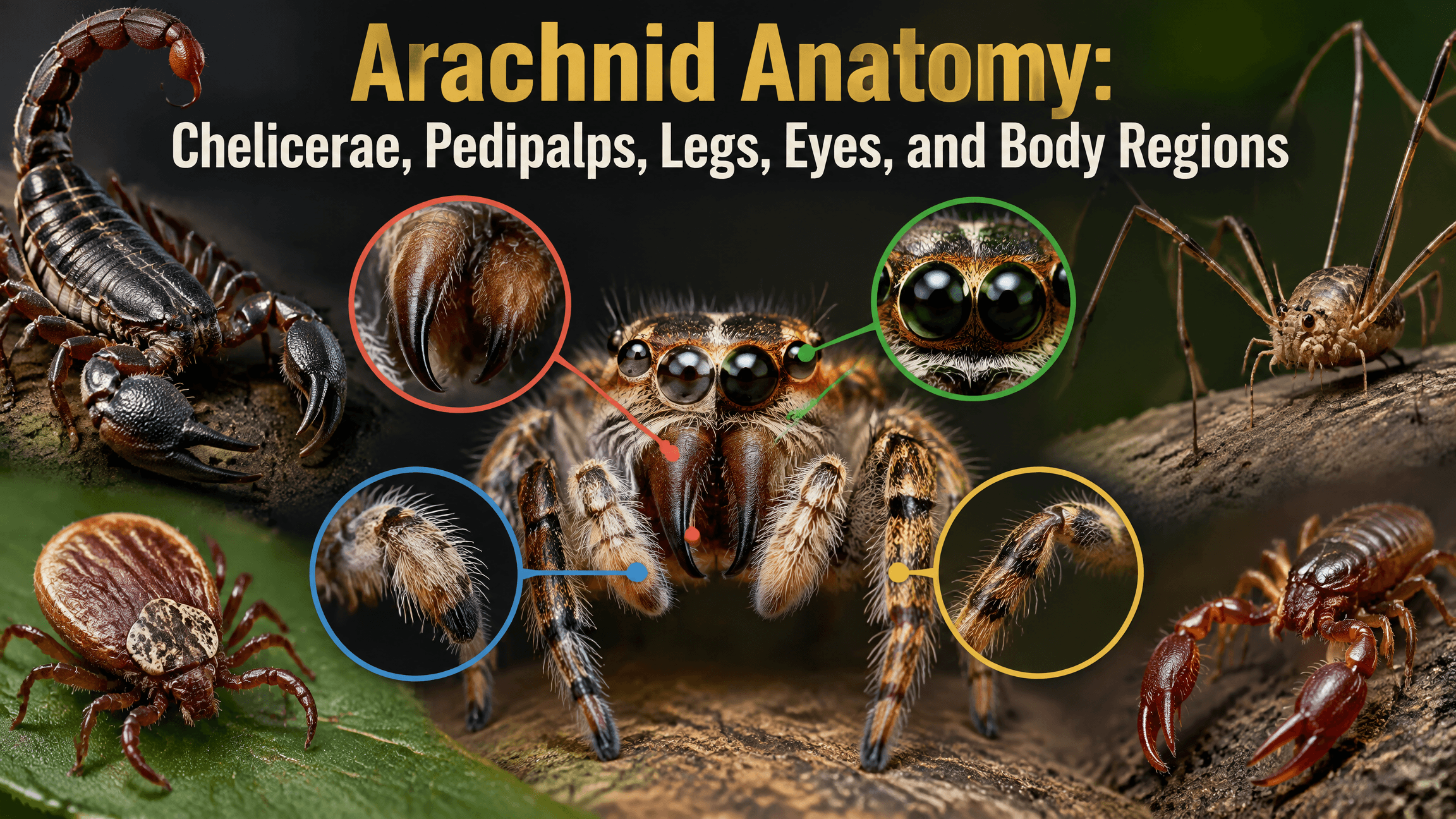
Arachnid anatomy follows a shared chelicerate plan, but that plan is far more flexible than the familiar spider silhouette suggests. Spiders, scorpions, ticks, mites, harvestmen, solifuges, pseudoscorpions, and whip spiders all possess the basic arachnid appendage pattern, yet those structures can look and function very differently from one group to another.
The most useful way to understand an arachnid body is to compare homologous structures, meaning body parts that share an evolutionary origin. Chelicerae can become spider fangs, small feeding appendages in scorpions, large mechanical jaws in solifuges, or part of the complex feeding apparatus of ticks. Pedipalps can be sensory structures, reproductive organs, grasping pincers, or raptorial prey-capture tools. Even the familiar idea of “two body sections and eight legs” needs exceptions and careful wording.
Quick Answer

Most adult or post-larval arachnids have a prosoma at the front that bears the chelicerae, pedipalps, and four pairs of walking legs, plus a posterior region commonly called the opisthosoma. However, the visible separation and shape of those regions vary greatly. The American Arachnological Society’s overview of arachnid groups shows how strongly the basic chelicerate plan has been modified among spiders, scorpions, mites, harvestmen, whip spiders, and other lineages. These structures are part of a broader arachnid body plan that has diversified in very different ways across Arachnida.
Some arachnids have conspicuous eyes, while others have few or none. Respiratory structures can include book lungs, tracheae, combinations of the two, or other arrangements in very small forms. The exoskeleton supports and protects the body, but the anatomy that distinguishes arachnid groups comes mainly from how the body regions and appendages are organized and specialized.
The Arachnid Body Plan Is Shared but Highly Variable
Prosoma and opisthosoma as comparative terms
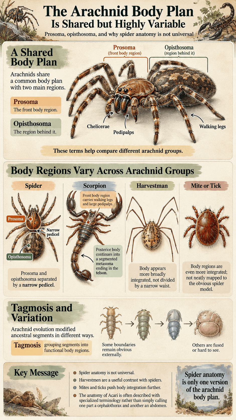
The front body region of an arachnid is usually called the prosoma. It carries the chelicerae, pedipalps, and walking legs. The region behind it is commonly called the opisthosoma. These terms are useful because they work across several arachnid groups without forcing every animal into the simpler “cephalothorax plus abdomen” language commonly used for spiders. Taken together, chelicerae, pedipalps, walking legs, and body organization contribute to the defining arachnid traits used in classification.
In a typical spider, the prosoma and opisthosoma are visibly separated by a narrow pedicel. That distinct waist makes the two regions easy to see. Scorpions are organized differently: the front body region carries the walking legs and large pedipalps, while the posterior body continues into a segmented metasoma ending in the telson. Harvestmen and many mites show much less obvious external separation between the major body regions.
Segmentation, fusion, and why spider anatomy is not universal
Arachnid evolution has modified ancestral segments in different ways. Some boundaries remain obvious externally, while others are fused or difficult to see. This process of grouping segments into functional body regions is called tagmosis.
Harvestmen are a useful contrast with spiders. In many familiar harvestmen, the main body appears broadly integrated rather than divided by a narrow waist. American Arachnological Society materials on Opiliones emphasize that harvestmen are a distinct arachnid lineage with an external body form that differs clearly from long-legged spiders.
Mites and ticks push body integration even further. Their traditional body subdivisions do not map neatly onto the obvious spider model, and many diagnostic structures are tiny. This is one reason the anatomy of Acari is often described using specialized terminology rather than simply calling one part a cephalothorax and another an abdomen.
Chelicerae: The First Feeding Appendages

Spider fang-bearing chelicerae
Chelicerae are the first pair of appendages in front of the mouth. In spiders, each chelicera typically includes a basal segment and a movable fang. The fang can deliver venom from associated glands in most spider lineages, although venom systems have been reduced or lost in some groups.
The spider chelicera is therefore both a feeding structure and part of a prey-subduing system. It is not simply a “tooth.” The appendage moves, grips, punctures, and helps position prey while the mouth takes in liquid food. The American Arachnological Society’s spider overview identifies fang-bearing chelicerae and abdominal silk organs among the defining features of Araneae.
Scorpion feeding chelicerae and separate venom stinger
Scorpion chelicerae are much smaller than their large pedipalps. They are used in handling and processing food near the mouth. The venom-delivery structure is elsewhere: at the end of the metasoma is the telson, which includes the venom apparatus and aculeus, or sting. The same structures can also become part of arachnid defenses through fangs, pincers, armor, silk, or other specializations.
This distinction prevents a common anatomy error. Scorpions do not inject venom through their chelicerae the way spiders usually do. Their large front pincers are also not chelicerae. Those pincers are modified pedipalps. The American Arachnological Society’s scorpion profile describes the combination of grasping pedipalps and the segmented metasoma ending in a venomous sting.
Tick mouthpart complex and hypostome
Tick feeding structures are highly modified for attachment to a host. The mouthpart complex includes chelicerae as well as a central structure called the hypostome. Depending on the tick group, the hypostome can carry backward-directed denticles that help maintain attachment while the animal feeds.
These structures do not look like a spider fang system, even though both spiders and ticks are chelicerates. The difference reflects very different feeding lifestyles. A spider usually uses its chelicerae to seize and penetrate prey, while a tick uses a specialized mouthpart assembly for prolonged ectoparasitic feeding.
Harvestman and solifuge chelicerae
Harvestmen have chelicerae used to grasp and process food, but they lack spider-type venom-delivery fangs. Because some harvestmen can take solid food particles, their feeding mechanics also differ from the strongly liquid-feeding pattern typical of many spiders.
Solifuges provide the opposite visual extreme. Their chelicerae can be very large and mechanically powerful, forming much of the front profile of the animal. Large chelicerae do not automatically mean venom. In most solifuges, mechanical processing is the important feature, and viral descriptions of “venomous camel spider jaws” confuse size with function.
Pedipalps: From Sensory Organs to Pincers and Raptorial Tools
Spiders
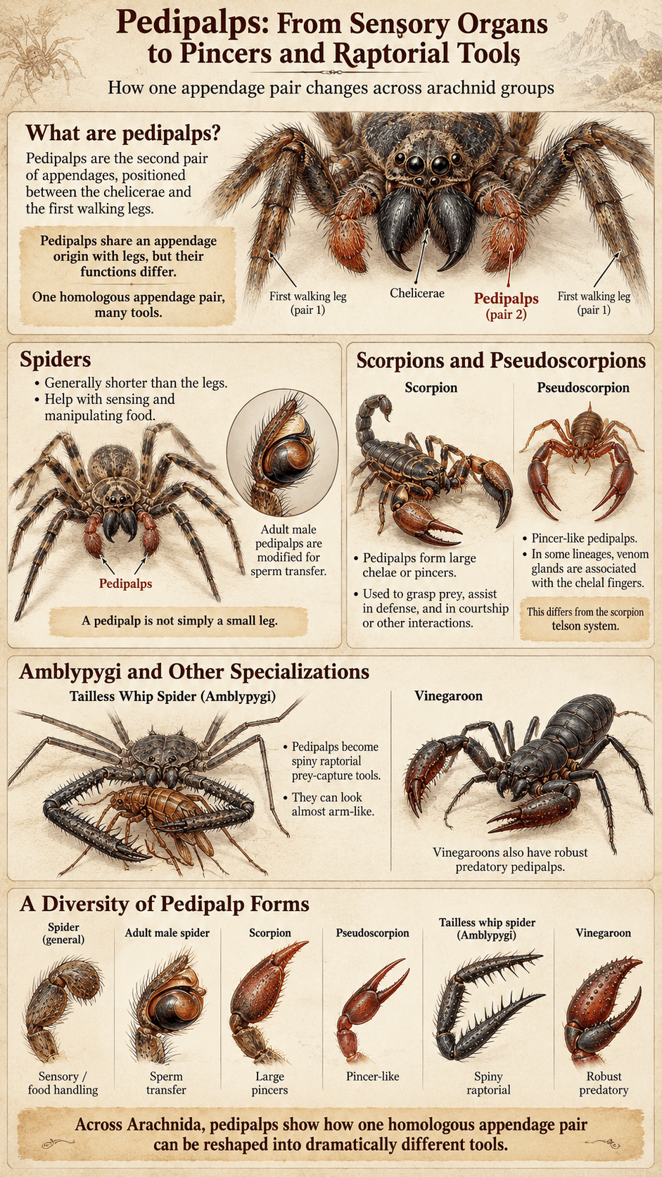
Spider pedipalps are the second pair of appendages, located between the chelicerae and the first walking legs. They are generally shorter than the legs and can help with sensing and manipulating food. In adult male spiders, the terminal parts of the pedipalps are modified into specialized structures used to transfer sperm during mating.
This is one of the clearest examples of a body part serving different roles across life and sex. A pedipalp is not simply a “small leg.” Its segments share an appendage origin with legs, but its function has been redirected toward sensory, feeding, and reproductive tasks.
Scorpions and pseudoscorpions
In scorpions, the pedipalps form the large chelae, or pincers, that dominate the front of the animal. They grasp prey, assist in defense, and are involved in courtship and other interactions. Their size and proportions differ among species, and pincer shape can reflect different mechanical strategies rather than a simple measure of venom strength or danger.
Pseudoscorpions also have prominent pincer-like pedipalps. In some lineages, venom glands are associated with the chelal fingers. That system differs from the scorpion telson and shows again how a shared appendage can be modified in different ways.
Amblypygi and other specializations
Tailless whip spiders, or amblypygids, turn the pedipalps into spiny raptorial structures that fold around prey. These appendages can look almost arm-like because they extend forward and carry rows of spines used in prey capture and defense.
Vinegaroons also have robust predatory pedipalps, although their overall body plan differs from Amblypygi. Across Arachnida, pedipalps demonstrate the same evolutionary theme repeatedly: one homologous appendage pair can be reshaped into dramatically different tools.
Walking Legs and Specialized Leg Functions
Typical four-pair post-larval pattern
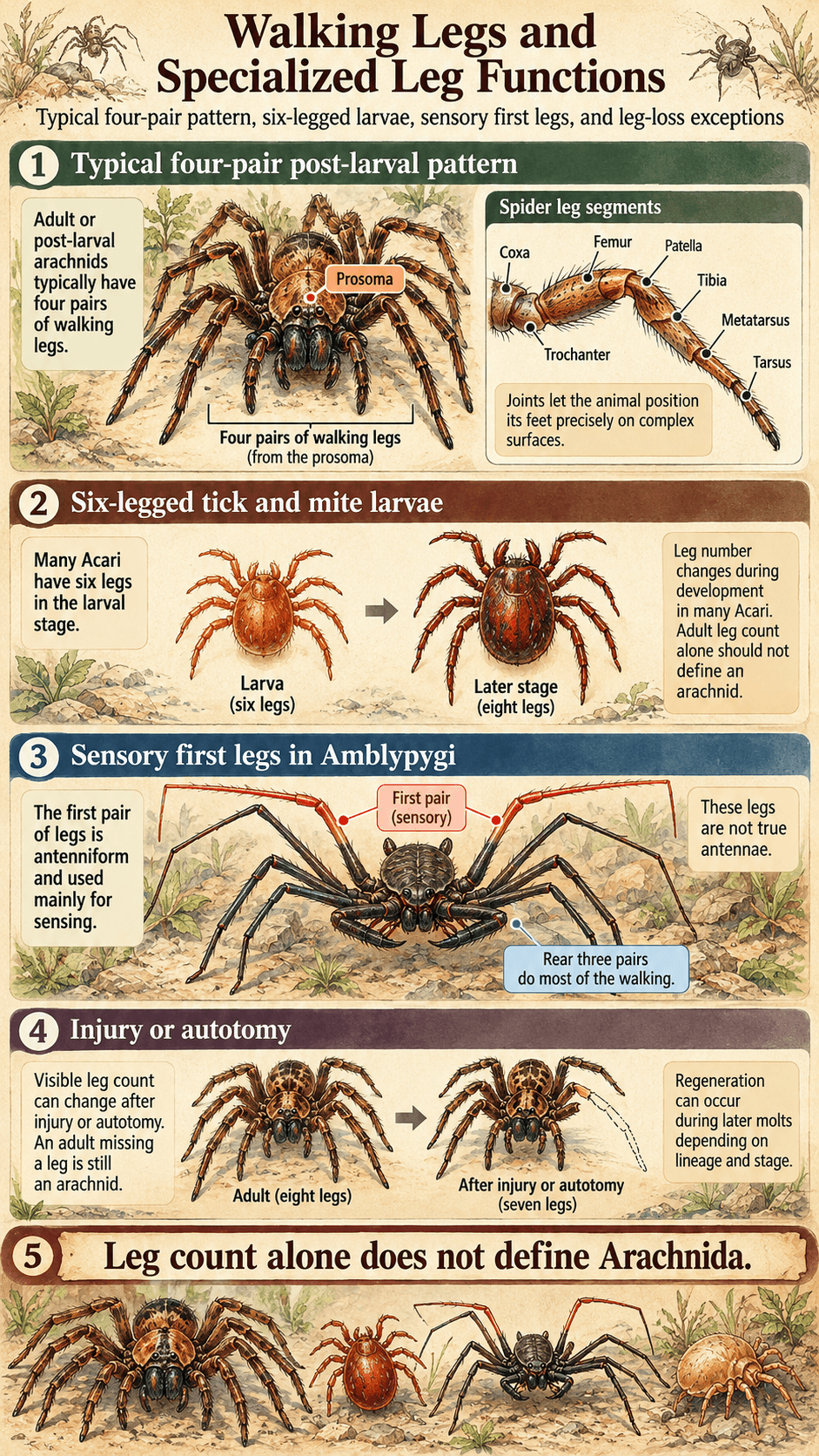
The familiar arachnid walking pattern is four pairs of legs attached to the prosoma. In many adult spiders, scorpions, harvestmen, and related groups, all eight serve mainly in locomotion, although individual legs also carry sensory hairs and receptors.
Each leg is divided into multiple articulated segments. The names and exact functional mechanics vary somewhat by group, but a spider leg, for example, includes segments such as the coxa, trochanter, femur, patella, tibia, metatarsus, and tarsus. Joints between these segments let the animal position its feet precisely on complex surfaces.
Six-legged tick and mite larvae
Leg number changes during development in many Acari. A typical tick larva emerges with three pairs of legs, giving it six legs rather than eight. After molting to the nymphal stage, it has four pairs. Many mites also have a six-legged larval stage, although developmental patterns across the group can be more varied.
The American Arachnological Society’s Acari overview emphasizes how diverse and taxonomically complex mites and ticks are. This developmental exception is one reason adult leg count alone should not be used as the definition of an arachnid.
Sensory first legs in Amblypygi and effects of injury or autotomy
Amblypygids appear to have eight long legs, but the first pair is not used primarily for ordinary walking. These extremely elongated appendages are antenniform, meaning antenna-like in function, and are packed with sensory structures. The animal normally walks on the rear three pairs.
The American Arachnological Society’s Amblypygi profile describes these first legs as flexible sensory appendages and identifies the spiny pedipalps as the major prey-capture structures. Calling the first legs true antennae would still be incorrect because arachnids do not possess insect antennae.
Visible leg count can also change after injury or autotomy, the controlled loss of an appendage. Some arachnids can regenerate lost leg tissue during later molts, but the ability depends on lineage, developmental stage, and the circumstances of loss. An adult missing a leg has not ceased to be an arachnid.
Eyes and Vision Structures
Spider eye-number and arrangement diversity
Many well-known spiders have eight simple eyes, but eight is not a universal spider number and certainly not a universal arachnid number. Eye arrangement differs among spider families, and some species have six, four, two, or no functional eyes. Cave-adapted spiders can show strong eye reduction.
Spider eyes are simple lens eyes rather than the compound eyes typical of many insects. Different eyes can be specialized for different tasks. Jumping spiders are famous for large forward-facing principal eyes and strong visual performance, while many web-building species rely heavily on vibration and touch even though they still possess eyes.
Scorpion, harvestman, tick, and mite differences
Scorpions usually have a pair of median eyes on top of the carapace and additional lateral eyes along the sides, although numbers vary among taxa. Their sensory world also depends heavily on nonvisual information from mechanoreceptors and specialized structures such as pectines.
Many harvestmen have a central ocularium, a raised area that bears a pair of eyes, although some lineages differ. Ticks and mites vary enormously: some possess eyes, some have simple light-sensitive structures, and others are effectively eyeless. Because Acari occupy habitats from exposed vegetation to soil, water, hosts, and caves, one eye plan could not describe the whole group accurately.
Cave adaptation and reduced eyes
Arachnids that spend their evolutionary history in caves may show reduced or absent eyes. Reduced pigmentation and elongated appendages can also occur, but these traits vary among lineages and should not be treated as a mandatory cave-animal package.
Eye loss is useful because it shows how anatomy responds to sensory priorities. In darkness, maintaining complex visual systems can offer little advantage, while touch, vibration, chemical detection, and long sensory appendages may become more important.
Mouthparts and Feeding Structures
How feeding anatomy differs among predators, parasites, and other diets
Arachnid feeding anatomy reflects diet. Spiders usually rely on fang-bearing chelicerae and extraoral digestion, releasing digestive fluids onto or into prey before ingesting liquefied material. Scorpions use chelicerae close to the mouth to tear and process food after prey has been subdued.
Harvestmen can process solid particles to a greater degree than spiders, while many mites have tiny piercing, scraping, cutting, or sucking structures adapted to plant tissue, fungi, detritus, small prey, or animal hosts. This diversity is one reason “arachnid mouthparts” is a broader concept than “fangs.”
Why tick feeding is not spider-like biting
A tick’s feeding apparatus works as a coordinated attachment system. Cheliceral components cut or manipulate tissue while the hypostome helps anchor the mouthparts. Salivary secretions interact with the feeding site, and the tick remains attached while taking a blood meal.
That prolonged ectoparasitic strategy differs from the rapid prey-capture bite of many spiders. Both use chelicerate-derived structures, but the anatomy has been reshaped for different biological problems.
Respiratory Anatomy
Book lungs
Book lungs are internal respiratory organs made of many thin, stacked lamellae that create a large surface area for gas exchange. Openings called spiracles connect the respiratory chamber with the outside. Scorpions use book lungs, and book lungs also occur in several spider and other tetrapulmonate lineages.
The name comes from the appearance of the layered plates, which resemble pages. Book lungs are not external gills and should not be imagined as lungs identical to those of mammals. They are arachnid respiratory organs with their own structure and evolutionary history.
Tracheal systems
Tracheae are tubes that carry gases through the body and open externally through spiracles. Many spiders possess tracheal systems, sometimes alongside book lungs. Mites and ticks show a broad range of respiratory arrangements, including tracheal systems in many groups and reduced or absent specialized respiratory structures in very small forms.
Body size matters because very small animals have short diffusion distances. Some tiny arachnids can rely more heavily on gas exchange across body surfaces or relatively simple internal pathways than a large scorpion could.
Mixed or reduced arrangements in different lineages
There is no single respiratory organ that defines Arachnida. Some spiders combine book lungs and tracheae, some lineages rely more strongly on one system, scorpions use book lungs, and Acari show extensive variation. Anatomy therefore has to be described group by group.
This variation also prevents a common myth: “all arachnids breathe through book lungs.” Book lungs are important, but they are only one part of a wider respiratory toolkit.
Circulatory and Nervous Systems at Overview Depth
Hemolymph and the open circulatory system
Arachnids have an open circulatory system. Their circulating fluid, called hemolymph, is pumped by a dorsal heart into body spaces rather than remaining entirely inside a closed network of veins and arteries like human blood.
Hemolymph moves nutrients, wastes, signaling molecules, and respiratory pigments where those are used. In spiders it also contributes to mechanical functions. Pressure in the body and legs helps extend certain leg joints, although the simple statement “spiders have hydraulic legs instead of muscles” is misleading.
Research in the Journal of Experimental Biology on spider leg mechanics shows that hydraulic extension works alongside muscular forces and that the balance depends on the joint and movement. The detailed biomechanics belong to locomotion, but anatomically the key point is that spiders use a hybrid system rather than replacing muscles altogether.
Central nervous organization and sensory integration
Arachnid nervous systems are concentrated in the prosoma, where nerve centers integrate signals from the eyes, mechanosensory hairs, chemical receptors, slit sensilla, pectines, Haller’s organ, and other structures depending on the lineage.
The relative arrangement differs among arachnids, especially in small or highly modified groups. What matters for a general anatomical comparison is that sensory structures distributed across appendages and body surfaces feed information into a centralized nervous system that coordinates movement and behavior.
Exoskeleton and Cuticle in Arachnids
What anatomy requires from cuticle and flexible membranes
Like other arthropods, arachnids are covered by a cuticle that forms the exoskeleton. Hardened plates can provide support and protection, while thinner flexible membranes at joints allow movement. Muscles attach internally to the exoskeleton and move appendage segments around their joints.
The balance between rigidity and flexibility is especially visible around leg joints, mouthparts, pedipalps, and the connections between major body regions. A completely rigid shell would prevent useful movement, so arachnid cuticle is organized into mechanically different regions.
Why body organization matters more here than exoskeleton mechanics
The presence of an exoskeleton is an arthropod-wide trait, not something unique to arachnids. What distinguishes arachnid anatomy more clearly is the arrangement of chelicerae, pedipalps, walking legs, body regions, sensory structures, and group-specific organs.
Molting is also essential because the rigid portions of the cuticle cannot expand indefinitely. Young arachnids replace the old cuticle as they grow. The detailed sequence of cuticle separation, shedding, expansion, and hardening is part of general arthropod molting rather than a feature that needs to be repeated as a separate arachnid anatomy system.
Spider, Scorpion, Tick, and Harvestman Anatomy Compared
Four body plans built from the same chelicerate foundation
| Feature | Spider | Scorpion | Tick | Harvestman |
|---|---|---|---|---|
| Body-region appearance | Distinct prosoma and opisthosoma joined by a narrow pedicel | Broad prosoma and opisthosoma continuing into a segmented metasoma | Highly integrated body outline with specialized feeding region | Body regions often appear broadly fused |
| Chelicerae | Usually fang-bearing and associated with venom | Small feeding appendages near the mouth | Part of a specialized host-feeding mouthpart complex | Grasp and process food without spider-style venom fangs |
| Pedipalps | Sensory and feeding roles; specialized for sperm transfer in adult males | Large grasping pincers | Reduced and incorporated into the feeding region | Leg-like or grasping depending on lineage |
| Venom delivery | Usually through cheliceral fangs | Through the telson and sting | No spider-style or scorpion-style venom apparatus | No spider-type venom-delivery fangs |
| Eye pattern | Variable, often multiple simple eyes | Median and lateral simple eyes in many species | Variable, including eyeless forms | Often a central pair on an ocularium, with variation |
| Respiration | Book lungs, tracheae, or combinations depending on lineage | Book lungs | Group-specific tracheal and other arrangements | Tracheal system in living harvestmen, with lineage-specific details |
The table is a comparison, not a diagnostic key. Each of these groups contains internal variation. Its value is showing how homologous parts have been reshaped. A chelicera is still a chelicera even when it looks nothing like a spider fang, and a pedipalp remains a pedipalp whether it is a spider palp or a scorpion pincer.
Common Anatomy Myths
All arachnids have two obvious body parts
Spiders make the two-region model easy to see because the prosoma and opisthosoma are joined by a narrow pedicel. That external pattern is not universal. Harvestmen can look broadly fused, and mites and ticks can have highly integrated body outlines.
The better approach is to learn the underlying organization and then examine how each lineage modifies it. Visible seams do not determine ancestry.
All arachnids have eight eyes
Eye number varies within Arachnida and even within spiders. Many spiders have eight, but others have fewer, and cave-adapted species can lack functional eyes. Scorpions, harvestmen, mites, and ticks follow different ocular patterns.
Eight eyes is therefore not an arachnid defining trait. It is one possible arrangement among many.
All arachnids have fangs, pincers, or book lungs
Spider-like fangs are specialized chelicerae and do not occur in every arachnid. Large pincers are modified pedipalps in scorpions and some other groups, not a universal feature. Book lungs occur in several major lineages but are absent from many others.
Arachnids are united by shared evolutionary architecture, not by one dramatic structure. Their anatomy makes more sense when the appendages are compared by origin first and by function second.
How Anatomy Shapes Movement, Sensing, and Feeding
Structures that determine how an arachnid moves
Leg construction, joint geometry, body mass, and attachment structures all influence locomotion. Spiders use muscular and hydraulic forces at different joints, scorpions carry a heavy posterior metasoma while walking, harvestmen can use very long legs to span uneven surfaces, and amblypygids reserve the first leg pair mainly for sensory exploration.
Anatomy creates possibilities and limits, but it does not dictate one movement style for an entire group. Closely related arachnids can run, climb, burrow, jump, or move slowly through litter depending on their proportions and habitat. Leg joints and their supporting structures also help explain the different forms of arachnid locomotion.
Sensory structures are distributed across the body
Eyes are only part of arachnid sensing. Fine hairs can detect touch and air movement, slit-like mechanoreceptors can respond to strain, scorpion pectines sample the substrate, ticks carry Haller’s organ on the first leg pair, and amblypygids use antenniform legs as mobile sensory arrays.
This distributed sensory anatomy explains why an arachnid with modest vision can still navigate, hunt, find mates, or locate hosts effectively. The body surface and appendages are active information-gathering systems.
Feeding structures reflect ecological strategy
A predator that grabs mobile prey, a parasite that remains attached to a host, and a mite that grazes fungi face different mechanical problems. Arachnid mouthparts have diversified accordingly. Chelicerae, pedipalps, hypostomes, and surrounding structures can pierce, hold, cut, crush, manipulate, or anchor depending on the lineage.
That connection between structure and ecology is the central lesson of arachnid anatomy. The same ancestral appendages can be remodeled so thoroughly that their common origin is easier to see through comparative anatomy than through appearance alone.
FAQ
What are chelicerae?
Chelicerae are the first pair of appendages in chelicerates, including arachnids. They are associated with feeding but take many forms. In spiders they bear the fangs, in scorpions they are small feeding appendages near the mouth, in solifuges they can be large mechanical jaws, and in ticks they form part of a specialized host-feeding apparatus.
What are pedipalps?
Pedipalps are the second pair of appendages, immediately behind the chelicerae. Their function differs strongly among arachnids. Spider pedipalps help with sensing and food handling and are modified for sperm transfer in adult males. Scorpion pedipalps form the large pincers, while amblypygid pedipalps are spiny prey-capture structures.
Do all arachnids have eight legs?
Four pairs of walking legs are typical in post-larval arachnids, but the statement needs qualification. Tick and many mite larvae have six legs, amblypygids use the first pair mainly as sensory appendages rather than ordinary walking legs, and injury or autotomy can reduce the visible number. Leg count alone does not define Arachnida.
Do all arachnids have eight eyes?
No. Eye number and organization vary widely. Many spiders have eight simple eyes, but some have six, four, two, or none. Scorpions have different median and lateral arrangements, harvestmen often have a central pair, and mites and ticks range from eyed to eyeless forms.
Do all arachnids have book lungs?
No. Book lungs occur in scorpions and several other arachnid lineages, including many spiders, but arachnid respiration is diverse. Tracheal systems occur in many groups, some spiders combine book lungs and tracheae, and very small arachnids can have reduced respiratory structures. Book lungs are important arachnid organs, not a universal defining trait.
Final Thoughts
Arachnid anatomy is easiest to understand as a flexible chelicerate framework rather than a checklist based on spiders. Chelicerae, pedipalps, walking legs, the prosoma, and the opisthosoma provide a shared foundation, but each lineage reshapes those parts for its own way of feeding, moving, sensing, reproducing, and surviving.
Spiders place venom-bearing fangs on the chelicerae, scorpions move venom delivery to a terminal sting and enlarge the pedipalps into pincers, ticks transform the mouth region for prolonged host attachment, harvestmen integrate the body regions differently, and amblypygids turn the first legs into elaborate sensory structures. Those contrasts are what make arachnid anatomy useful: the differences reveal how one evolutionary body plan can support an extraordinary range of lives.

Ethan Walker is the founder and research editor of Animal Fact Central. He creates and reviews educational animal facts content using trusted wildlife, pet care, and science-based sources. His work focuses on making animal behavior, adaptations, habitats, and species facts clear, accurate, and engaging for everyday readers.
Read More Details About Ethan Walker: https://animalfactcentral.com/ethan-walker/