

Insect anatomy follows a remarkably consistent plan even though insects range from tiny parasitic wasps to heavy-bodied beetles, delicate moths, grasshoppers, dragonflies, and countless other forms. A typical adult insect has three main body regions, the head, thorax, and abdomen, plus three pairs of jointed legs, one pair of antennae, and an external skeleton. Wings may be present, modified, reduced, or absent.
That basic layout is useful because each region is specialized. The head concentrates sensory organs and mouthparts. The thorax carries all six adult legs and, when present, the wings. The abdomen contains much of the digestive, reproductive, and respiratory machinery. The Smithsonian’s overview of insect body traits highlights this three-part plan and the thoracic attachment of legs and wings.
Looking beyond the outside reveals an equally distinctive internal design. Insects do not have lungs or a closed network of arteries and veins like mammals. Instead, most use branching air tubes to deliver oxygen through the body, and they circulate hemolymph through an open body cavity. Their body wall is also more than a rigid shell. It combines hard plates, flexible membranes, living tissue, sensory structures, and attachment points for muscles.
Quick Answer

The insect body plan in one labeled framework
The easiest way to read an insect’s body is from front to back. The head bears the antennae, eyes, and mouthparts. Behind it, the thorax is divided into three segments and carries one pair of adult legs on each segment. The last two thoracic segments are also the usual attachment points for wings. The abdomen follows behind and contains most of the gut, reproductive organs, and many spiracles, which are openings associated with the respiratory system.
These regions are not simple boxes. Each is built from segments and hardened plates joined by softer areas of cuticle. Appendages are jointed, and internal ridges of the exoskeleton provide places for muscles to attach. That combination gives an insect both protection and mobility.
External anatomy versus internal systems
External anatomy includes visible features such as antennae, eyes, mouthparts, legs, wings, abdominal segments, and the outer body wall. Internal anatomy includes the digestive tract, tracheal system, hemolymph and circulatory organs, nervous system, reproductive organs, muscles, glands, and other tissues.
The distinction is useful, but the two levels constantly interact. A spiracle is an external opening connected to internal tracheae. A sensory hair projects through the body surface but is linked to living receptor cells and nerves. A leg looks like an external appendage, yet it moves because muscles pull on internal parts of the exoskeleton. Insects function as integrated bodies, not as hard shells containing unrelated organs.
Head: Sensing and Feeding

Antennae and major sensory roles
An insect’s single pair of antennae attaches to the head. Antennae vary greatly in length, shape, and surface structure, but they are not merely “noses.” Depending on the species, receptors on the antennae can detect airborne chemicals, touch, air movement, humidity, temperature, and other physical or chemical cues.
The antenna itself is segmented and movable. Different groups have evolved threadlike, clubbed, feathery, elbowed, or otherwise specialized antennae. These shapes often reflect how an insect samples its surroundings, but appearance alone does not tell you everything the antenna can detect.
Compound eyes and ocelli
Many adult insects have a pair of compound eyes made of repeated visual units called ommatidia. Information from those units is processed by the nervous system into useful visual signals. It is misleading to imagine a compound eye as showing the insect thousands of completely separate little pictures.
Some insects also have simple eyes called ocelli. Ocelli do not function like tiny versions of compound eyes. They can contribute to detecting changes in light intensity, orientation, and flight control in particular groups. The exact number and role vary among insects and life stages.
Mouthparts as modified homologous structures
Insect mouthparts can look radically different, yet many are modifications of the same basic sets of structures. Mandibles, maxillae, the labium, and other components can be enlarged, reduced, lengthened, fused, or reorganized to handle different foods.
This is why a grasshopper can chew leaves, a true bug can pierce tissue and draw up fluids, a butterfly can take in liquid through a long proboscis, and a house fly can sponge liquid food even though all are insects. Mouthpart anatomy provides one of the clearest examples of evolution modifying a shared structural framework for different functions.
Thorax: Legs, Wings, and Movement
Prothorax, mesothorax, and metathorax
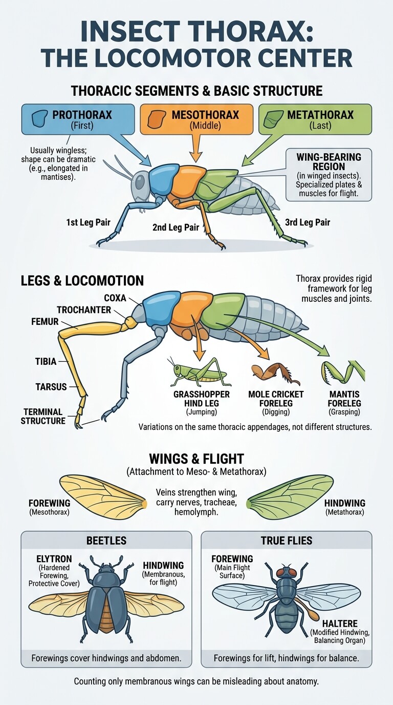
The insect thorax consists of three segments called the prothorax, mesothorax, and metathorax. These names simply mean the first, middle, and last thoracic segments. One pair of adult legs attaches to each, giving an insect three pairs in total.
In winged insects, the mesothorax and metathorax form the main wing-bearing region. Their skeletal plates and muscles can be highly specialized for flight. The prothorax usually does not carry wings in living adult insects, although its shape can be dramatic, as in mantises with an elongated first thoracic segment.
Why all three adult leg pairs attach to the thorax
Adult insect legs arise from the thorax because this region is the main locomotor center. Each leg has a series of jointed sections, commonly including the coxa, trochanter, femur, tibia, tarsus, and terminal structures. The exact shape and proportion of these parts differ greatly among groups.
The thorax provides the rigid framework needed for leg muscles and joints. It also allows the three leg pairs to specialize without changing the underlying body plan. A grasshopper’s enlarged hind legs, a mole cricket’s digging forelegs, and a mantis’s grasping forelegs are all variations on thoracic appendages rather than completely different structures.
Wing attachment and the basic anatomy of forewings and hindwings
When insects have wings, they attach to the second and third thoracic segments. These are commonly called forewings and hindwings according to position. In many groups both pairs contribute directly to flight. In others, one pair is heavily modified.
Beetle forewings form hardened elytra that cover the more delicate hindwings and much of the abdomen. In true flies, the forewings provide the main flight surfaces while the hindwings are modified into halteres, small balancing organs. These examples show why counting only obvious membranous wings can give the wrong impression about insect anatomy.
Wing surfaces contain veins that strengthen the wing and can carry nerves, tracheae, and hemolymph. The detailed mechanics of wing flexion, wingbeat, aerodynamic force, and flight control are separate questions from basic anatomy, but all begin with structures rooted in the thorax.
Abdomen: Respiration, Digestion, Reproduction, and Flexible Segmentation
Abdominal segmentation and flexible membranes
The insect abdomen is visibly segmented in many species, although some segments may be reduced, fused, or hidden. Hardened plates protect the body, while flexible membranes between them allow bending, expansion, breathing movements, feeding-related swelling, egg development, and other changes in shape.
This flexible construction is important because a completely rigid tube would be a poor abdomen. Female insects that develop large numbers of eggs, blood-feeding insects that take a large meal, and insects that ventilate the respiratory system with abdominal pumping all depend on controlled flexibility.
Spiracle distribution and internal organ context
Spiracles are valve-like openings that connect the outside air to the tracheal system in many insects. They usually occur along the sides of thoracic and abdominal segments, although number, placement, and function vary widely. They are not simply open holes. Many insects can regulate them, helping balance gas exchange with water loss.
Much of the digestive tract runs through the abdomen, along with excretory structures, reproductive organs, fat body, muscles, and the dorsal vessel associated with circulation. These organs share the body cavity rather than being separated into the same arrangement of closed compartments found in vertebrates.
Reproductive structures including the ovipositor
Reproductive structures differ enormously among insect groups. Females of many species have an ovipositor, an egg-laying structure derived from abdominal appendages. It may be short and inconspicuous or highly specialized for placing eggs in soil, plant tissue, hosts, wood, or other sites.
In some hymenopterans, components of the ovipositor system have been modified into a sting used in defense or prey handling. That does not mean every female insect with an ovipositor can sting, or that every hymenopteran possesses a functional sting.
The Insect Exoskeleton and Cuticle
Cuticle, living epidermis, sclerites, and flexible membranes
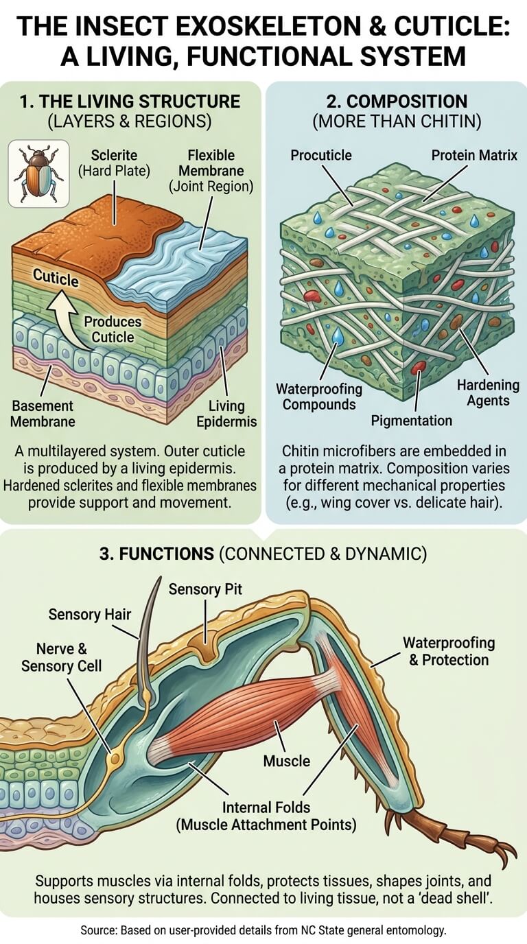
The insect exoskeleton is often described as armor, but that description is incomplete. The outer cuticle is produced by a living epidermis underneath it. Hardened regions form plates called sclerites, while flexible membranes remain between many plates and around joints. This mixed construction provides both support and movement.
NC State’s general entomology treatment of the insect exoskeleton describes the integument as a multilayered system that includes cuticle, epidermis, and basement membrane. It also explains how hardened and flexible regions can differ even within the same insect.
Chitin plus proteins rather than chitin alone
It is common to hear that insect exoskeletons are “made of chitin.” Chitin is important, but that statement is too simple. In the procuticle, chitin microfibers are embedded in a protein matrix. Other compounds contribute to the outer layers, waterproofing, pigmentation, and hardening.
Different areas of the body can have different mechanical properties. A beetle’s hardened wing cover, a flexible joint membrane, and a delicate sensory hair all involve cuticular material, but they are not identical in composition or stiffness.
How the exoskeleton supports joints, muscles, protection, and sensory structures
The exoskeleton provides attachment surfaces for muscles, protects internal tissues, reduces water loss, shapes joints, and supports sensory structures. Internal folds or ridges of the body wall can act as bracing and muscle attachment points, allowing powerful movement without an internal bony skeleton.
The body surface also carries hairs, bristles, scales, pits, and other structures that may have sensory or protective functions. Because these structures are connected to living tissue, the exoskeleton should not be imagined as a dead shell that the insect merely wears.
Six Legs and Their Specializations
Walking and running legs
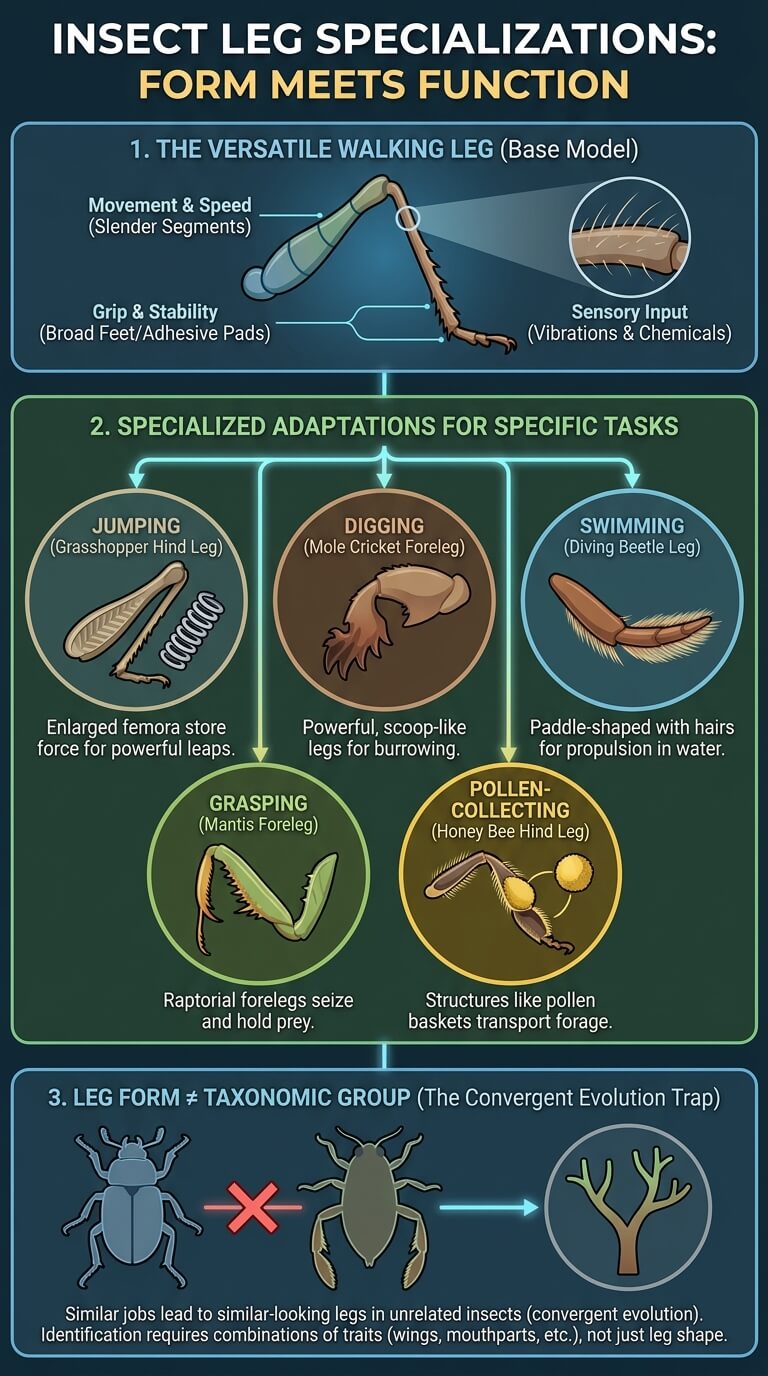
The basic insect leg is a chain of jointed segments, but proportions differ according to how an insect moves. In many ground-running insects, long legs and relatively slender segments help produce rapid strides. In other species, broader feet or adhesive structures improve grip on leaves, bark, smooth surfaces, or prey.
Even seemingly ordinary walking legs can carry dense sensory hairs and chemical receptors. A leg can therefore help an insect move, test surfaces, detect vibrations, and in some species sample chemicals at the same time.
Jumping, digging, swimming, grasping, and pollen-collecting modifications
Specialized insect legs show how much one body plan can be reshaped. Grasshoppers and crickets often have enlarged hind femora that store and transmit force for jumping. Mole crickets have broad, powerful forelegs for digging. Diving beetles can have flattened, fringed legs that work like paddles.
Mantises have raptorial forelegs designed to seize prey. Honey bee workers possess specialized structures on the hind legs that help handle and transport pollen. Aquatic insects may use rows of hairs to increase the effective surface area of swimming legs.
Why leg form does not define a taxonomic group by itself
Similar jobs can lead unrelated insects to evolve similar-looking leg modifications. A swimming leg is not evidence that all insects with paddle-shaped legs belong to the same order. The same caution applies to grasping, digging, or jumping legs.
Entomologists identify insects by combinations of traits, including wings, mouthparts, antennae, development, genital structures, and many other features. Leg shape is useful, but it is one clue among many.
Mouthpart Designs and What They Can Do
Chewing
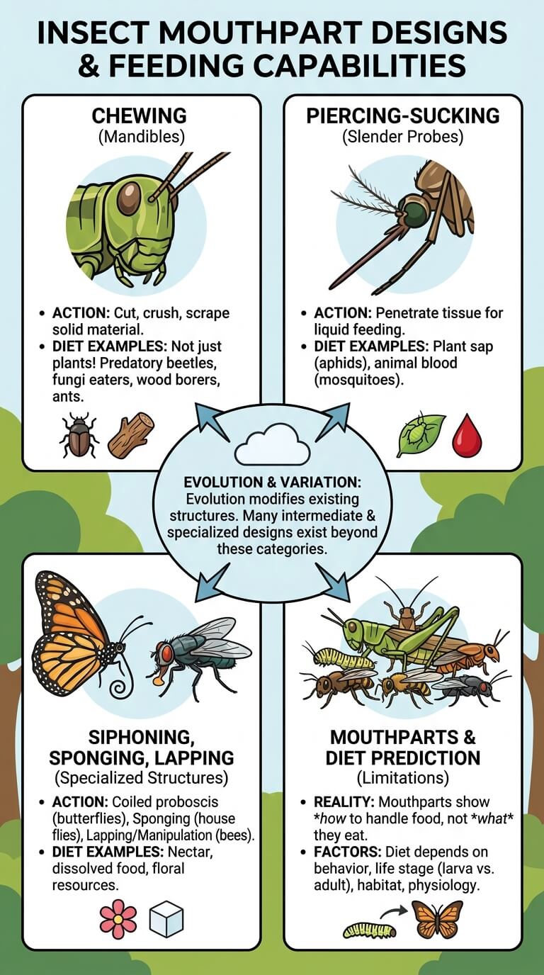
Chewing mouthparts use mandibles to cut, crush, scrape, or manipulate solid material. Grasshoppers, many beetles, cockroaches, ants, and numerous larvae provide familiar examples, although what they actually eat varies greatly.
Chewing should not be treated as a synonym for plant feeding. Predatory beetles also chew, as do insects that eat fungi, seeds, wood-associated material, animal matter, or mixed diets.
Piercing-sucking
Piercing-sucking mouthparts contain slender components that penetrate tissue and allow liquid feeding. Aphids and many other hemipterans use specialized mouthparts to access plant fluids. Mosquitoes use a very different piercing apparatus, and feeding differs between sexes and among life stages.
The shared label describes a mechanical strategy, not one universal diet. Plant sap, animal blood, and other liquid resources require different behaviors, enzymes, and physiological adaptations.
Siphoning, sponging, lapping, and chewing-lapping
Adult butterflies and many moths have a coiled proboscis adapted for taking in liquids. House flies use sponging mouthparts to collect liquid or dissolved food. Bees combine several mouthpart structures in ways that allow lapping and manipulation of floral resources and other materials.
These familiar categories are useful teaching tools, but real insect mouthparts show many intermediate and specialized designs. Evolution modifies existing structures rather than building each feeding apparatus from an entirely new set of parts.
Why mouthpart type does not perfectly predict diet
Mouthparts tell you how an insect can handle food, but not always exactly what it eats. Closely related species can use similar structures on different resources, and an insect’s diet can change dramatically between larval and adult stages.
For that reason, anatomy and feeding ecology should be kept distinct. Mouthpart design sets mechanical possibilities and constraints, while actual diet also depends on behavior, habitat, life stage, digestive physiology, and available foods.
How Insects Breathe
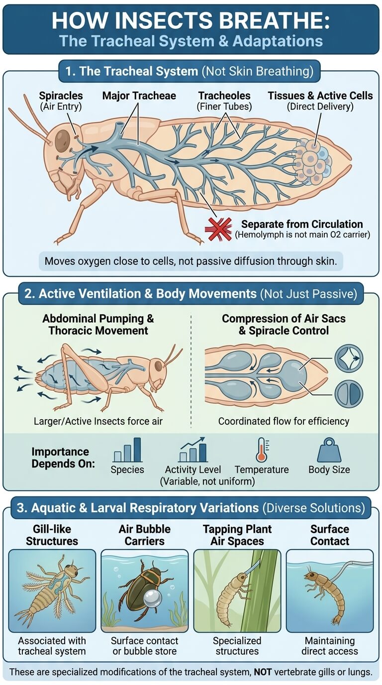
Spiracles and the tracheal system
Most insects exchange respiratory gases through a branching tracheal system rather than lungs. Air enters through spiracles and moves into larger tracheae that divide repeatedly into finer tubes reaching tissues. This arrangement brings oxygen close to active cells without relying on hemolymph as the main oxygen carrier.
The NC State overview of insect respiration describes the tracheal network as a system separate from circulation. This is why saying that insects “breathe through their skin” is inaccurate. Their body surface contains specialized openings connected to internal air tubes.
Active ventilation and body movements in some insects
Diffusion is important at small distances, but insect respiration is not always a completely passive process. Many larger or highly active insects can move air using abdominal pumping, thoracic movements, compression of air sacs, or coordinated opening and closing of spiracles.
The importance of these mechanisms depends on species, activity level, temperature, and body size. It is therefore safer to describe insect ventilation as variable rather than claim that oxygen simply drifts through every insect in the same way.
Aquatic and larval respiratory variations
Aquatic insects show especially diverse solutions. Some larvae or nymphs use external or internal gill-like structures associated with the tracheal system. Others carry air bubbles, maintain contact with the surface, tap plant air spaces, or use specialized structures to exchange gases underwater.
These variations do not make them vertebrate-style gills or lungs. They are modifications of insect respiratory biology suited to habitats where direct access to atmospheric air may be limited.
Hemolymph and Circulation
Open circulatory system at a general-reader level
Insects have an open circulatory system. Their circulating fluid, called hemolymph, is not confined to a closed network of arteries, veins, and capillaries. A dorsal vessel helps move hemolymph, which then circulates through the main body cavity and around organs.
Hemolymph transports nutrients, hormones, salts, wastes, and immune cells, and it can contribute to hydraulic movements and wound sealing. The NC State guide to insect circulation explains how this open system differs from vertebrate circulation.
Why hemolymph usually does not transport oxygen like vertebrate blood
Human blood relies heavily on red blood cells and hemoglobin to move oxygen. Most insects solve the oxygen-delivery problem differently because the tracheal system carries gases directly toward tissues. As a result, hemolymph usually does not play the same respiratory role as vertebrate blood.
This distinction also explains why insect hemolymph need not be red. It may be clear, pale, yellowish, greenish, or another color depending on pigments, diet, physiology, and species.
Specialized exceptions with respiratory pigments
There are exceptions to the general rule. Some aquatic insect larvae, including certain midges, possess hemoglobin or other respiratory adaptations that help them live where oxygen availability can be low. These cases are reminders that insect physiology is diverse.
The best general statement is therefore that the tracheal system handles most respiratory gas transport in most insects, while specialized lineages may add other mechanisms.
Digestive and Nervous Systems
Foregut, midgut, and hindgut at overview depth
The insect digestive tract is commonly divided into foregut, midgut, and hindgut. Food enters through the mouth and foregut, chemical digestion and nutrient absorption occur largely in the midgut, and the hindgut plays important roles in handling waste and recovering water and salts.
NC State’s insect digestive system overview also describes associated structures such as salivary glands and Malpighian tubules. The exact shape of each region varies greatly with diet and insect group.
Brain, ventral nerve cord, and distributed neural control
An insect nervous system includes a brain in the head, additional ganglia, and a ventral nerve cord that extends through the body. Ganglia are groups of nerve cells that process information and help control local movements and organ functions.
This arrangement means control is more distributed than in a vertebrate nervous system. Thoracic ganglia, for example, are closely involved in leg and wing movement, while abdominal ganglia help coordinate abdominal structures. The NC State description of the insect nervous system shows how the brain and segmental ganglia work together rather than treating the brain as the only control center.
Common Anatomy Myths
Do insects breathe through their skin?
Not in the ordinary sense of that phrase. Most insects use spiracles connected to an internal tracheal system. Gas exchange ultimately occurs across very thin surfaces near tissues, but the organized tube network is the key feature. Describing this as simple skin breathing hides the anatomy that makes insect respiration distinctive.
Is the exoskeleton a dead shell?
No. The outermost cuticle itself is secreted material, but it is produced by living epidermal cells and integrated with muscles, glands, nerves, and sensory receptors. Flexible membranes, joints, and sensory structures are all part of the functional body wall.
An insect also changes this system as it grows. Molting involves separating from and replacing old cuticular layers, then expanding and hardening new ones. The developmental details belong to insect growth and life-cycle biology, but they reinforce the point that the exoskeleton is a dynamic biological structure.
Is insect blood the same as vertebrate blood?
No. The fluid is called hemolymph, and its circulation, cell types, and major functions differ from vertebrate blood. In most insects, oxygen delivery is handled mainly by the tracheal system rather than by red blood cells circulating through closed vessels.
Calling hemolymph “insect blood” can be a useful shortcut, but it should not imply identical anatomy or physiology.
How Anatomy Shapes Flight, Sensing, and Feeding
Wing structure sets the stage for insect flight
The thorax contains the skeletal framework and muscles that make flight possible, and the wings themselves are flexible structures supported by veins. How those structures generate lift, maneuver, fold, couple, or function as modified organs requires a closer look at insect flight mechanics.
Basic anatomy supplies the parts. Flight biology explains how those parts move together in real time.
Sensory structures turn the body surface into an information system
Antennae, compound eyes, ocelli, sensory hairs, vibration-sensitive organs, and chemical receptors all begin as anatomical structures, but their function depends on physiology and neural processing. An antenna is therefore more than an appendage, just as a compound eye is more than a patterned surface.
Studying sensory biology reveals how insects use those structures to find food, mates, shelter, hosts, and escape routes.
Mouthpart anatomy creates different feeding possibilities
Chewing, piercing, siphoning, sponging, and lapping structures place different mechanical limits on how an insect can obtain food. Yet diet cannot be predicted from anatomy alone. Behavior, digestive enzymes, microbial partners, life stage, and habitat can all shape what a species actually consumes.
The mouthparts are therefore a starting point for understanding feeding, not a complete menu.
FAQ
Do insects have bones?
Insects do not have an internal bony skeleton like mammals, birds, reptiles, amphibians, or fish. Their main supportive framework is an exoskeleton on the outside of the body. Internal ridges and folds of that exoskeleton also provide attachment points for muscles.
The absence of bones does not mean an insect lacks rigid support. Hardened cuticular plates can be extremely effective at supporting the body while remaining light enough for small-bodied locomotion and, in many groups, flight.
Do insects have lungs?
Most insects do not have lungs. They use a tracheal respiratory system made of branching tubes that connect to the outside through spiracles. Fine branches bring oxygen close to tissues and help remove carbon dioxide.
Aquatic insects may modify this system in several ways, so the details vary, but vertebrate-style lungs are not part of the standard insect body plan.
Where are an insect’s legs attached?
All three pairs of adult insect legs attach to the thorax. One pair belongs to each of the three thoracic segments: the prothorax, mesothorax, and metathorax.
This is one of the most useful features for recognizing insect anatomy. Appendages on the abdomen may have other functions, but they are not additional adult walking-leg pairs.
Do insects have blood?
Insects have a circulating fluid called hemolymph. It transports nutrients, hormones, salts, wastes, and immune components and helps with several hydraulic functions. Because insects have an open circulatory system, hemolymph is not restricted to a vertebrate-style network of arteries, veins, and capillaries.
In most insects, hemolymph also does not carry most of the oxygen. The tracheal respiratory system handles that job directly, with some specialized exceptions.
Final Thoughts
Insect anatomy makes more sense when the body is viewed as an integrated system. The head concentrates sensing and feeding structures, the thorax carries the six legs and wing machinery, and the abdomen houses much of the digestive, reproductive, and respiratory equipment. Around all of it, the exoskeleton combines hard sclerites with flexible membranes, living epidermis, joints, muscle attachments, and sensory structures.
Inside, branching tracheae deliver gases, hemolymph circulates through an open body cavity, the digestive tract is organized into major functional regions, and a brain plus ventral nerve cord coordinates activity through distributed ganglia. That combination helps explain why insects can share one recognizable body plan while evolving such different ways to walk, fly, feed, sense, and survive.

Ethan Walker is the founder and research editor of Animal Fact Central. He creates and reviews educational animal facts content using trusted wildlife, pet care, and science-based sources. His work focuses on making animal behavior, adaptations, habitats, and species facts clear, accurate, and engaging for everyday readers.
Read More Details About Ethan Walker: https://animalfactcentral.com/ethan-walker/